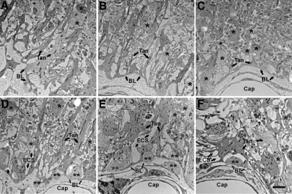
Figure 3.
Electron microscopic images of the pericapillary zone of the lateral median eminence are shown. A–C are from one representative vehicle-treated young rat with micrographs taken from most lateral (A) to medial (C) along the pericapillary zone. A, At the most lateral end of the median eminence, a few GnRH-immunopositive terminals (not identifiable at this magnification) are found. B, Moving more medially, the tanycyte processes gradually become organized and linear. C, In the most medial portion of the median eminence, a large number of neuroterminals are present, although relatively few GnRH-immunopositive terminals are found. Portal capillaries are covered by tanycytic endfeet, but few elongated tanycyte processes are found. D–F are representative data from three representative vehicle-treated rats: young (D), middle-aged (E), and old (F). Images were taken at a similar level of the median eminence pericapillary zone as that shown in B. In D–F, neuroterminals (single black asterisk), extracellular space (ECS; arrows), and the portal capillary (Cap) basal lamina (BL; arrows) can be seen. Similar profiles were seen for comparable estradiol-treated rats at each age (not shown). The neuroterminals apposed to the basal lamina (double black asterisks) have few large dense-core vesicles. The extracellular space (ECS) with low electron density surrounds neuronal and glial elements. Scale bar, 2 μm.

