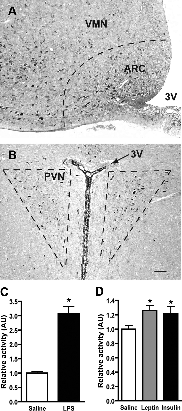
Figure 1.

Localization of aPKC in hypothalamus and response to LPS, leptin, and insulin. Representative images from frozen sections of perfused adult male rat brain stained with a polyclonal aPKC antibody. A, In the midcaudal region of the ventral hypothalamus, aPKC expression is largely restricted to neurons of the arcuate nucleus (ARC). Staining is also observed in the ventrolateral portion of the neighboring ventromedial nucleus (VMN). B, More restricted expression is seen in the PVN within the medial parvocellular region. Scale bar, 100 μm for both images. C, After a 4-h fast, rats (n = 9/group) were injected ip with saline or LPS (50 μg/kg) followed 2 h later by the animals being killed. In vitro aPKC kinase activity was determined on mediobasal hypothalamus extracts immunoprecipitated with a polyclonal aPKC antibody. Data (mean + sem) was normalized to saline-injected controls. *, P < 0.001. D, After a 4-h fast, 3V-cannulated rats (n = 8–12/group) were injected icv with saline, leptin (10 μg), or insulin (10 mU) followed by the animals being euthanized at 30, 45, and 20 min, respectively. aPKC kinase activity was determined as in C. *, P < 0.05 relative to saline control. AU, Arbitrary units.
