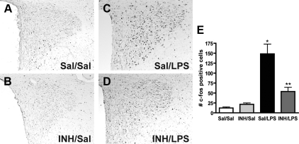
Figure 2.
Role of aPKC activity in LPS induction of c-fos in the PVN. A–D, Representative c-fos immunohistochemistry of PVN-containing sections from rats pretreated with icv saline (Sal) or INH (2 nmol) followed by ip saline or LPS (100 μg/kg). A and B, Both Sal/Sal and INH/Sal show minimal fos immunoreactivity. C, Sal/LPS animals show the expected increase in intensity and number of stained cells throughout the PVN. D, INH/LPS animals have less fos-positive neurons in the PVN, mostly due to reduction in the medial parvocellular group. E, Quantification of c-fos immunohistochemistry. Bars, mean + se of cell counts averaged from eight sections through the PVN in at least four animals/group. *, P < 0.05 vs. Sal/Sal and LPS/INH; **, P < 0.05 vs. Sal/Sal and Sal/LPS.

