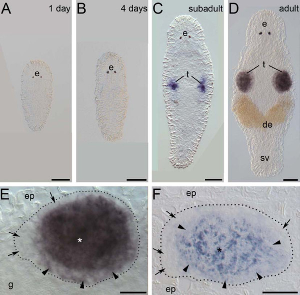
Figure 2.
Expression pattern of melav2 mRNA. (A, B) In 1-day old hatchlings (A) and 4-days old juveniles (B) no melav2 signal was detected. (C) Subadult worms. The signal was present in several cells of the developing testes (t). (D) Mature adult worms. Strong expression was detected in testes (t), but not in the seminal vesicle (sv). (E) Magnified image of testis of a mature adult worm (D). Cells on the edge of testis had no or only weak melav2 expression (arrow and arrowhead, respectively). Strong expression was detected mainly in the centre of testis (asterisk). (F) Sagittal section of the testis after whole mount in situ hybridization. The head is left and the dorsal side is up. Spermatogonia, and probably also spermatocyte I were melav2 negative (arrow). Weak signal was detected in spermatocyte II (arrowhead) and strong expression was detected in spermatids (asterisk). e, eyes; de, developing eggs; ep, epidermis; g, gut. Scale bars: A-D, 50 μm; E, F, 20 μm.

