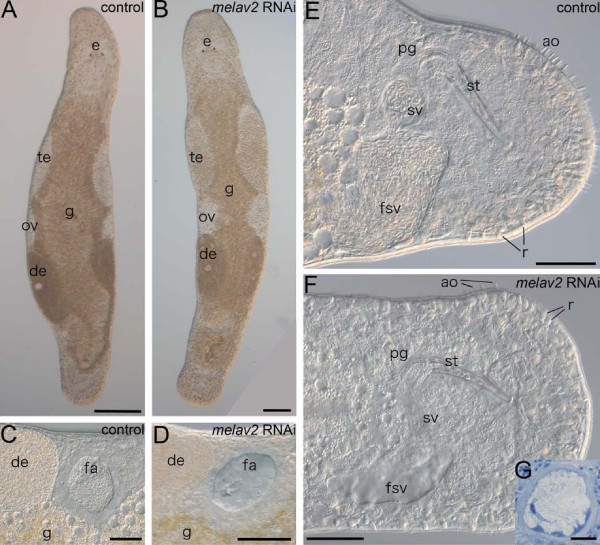
Figure 3.
Comparison of the overall morphology of control and melav2 RNAi treated M. lignano. (A, B) The overall of morphology of control animals (A) was comparable with that of melav2 RNAi treated animals (B) except for the morphology of the testis (te). Eyes (e), gut (g), ovaries (ov), developing eggs (de), and copulatory stylet (st) were not affected by the melav2 RNAi treatment. (C, D) The female antrum (fa) was filled with received sperm in the control animals (C), while no sperm was present in the melav2 RNAi treated animals (D). (E, F) In the control animals (E), the false seminal vesicle (fsv) and the seminal vesicle (sv) were filled with sperm, while no sperm was usually found there in the melav2 RNAi treated animals (F). The copulatory stylet (st), prostate glands (pg), rhabdites (r), gut (g) and adhesive organs (ao) were not affected by the melav2 RNAi treatment. Note that only a few adhesive organs are in the focal plane in this picture. (G) In a few cases, the melav2 RNAi treated animals had some aberrant spermatids in the seminal vesicle (G), suggesting that the vas deference was connected to the testis normally. Scale bars: A, B, 100 μm; C-F, 50 μm; G, 10 μm.

