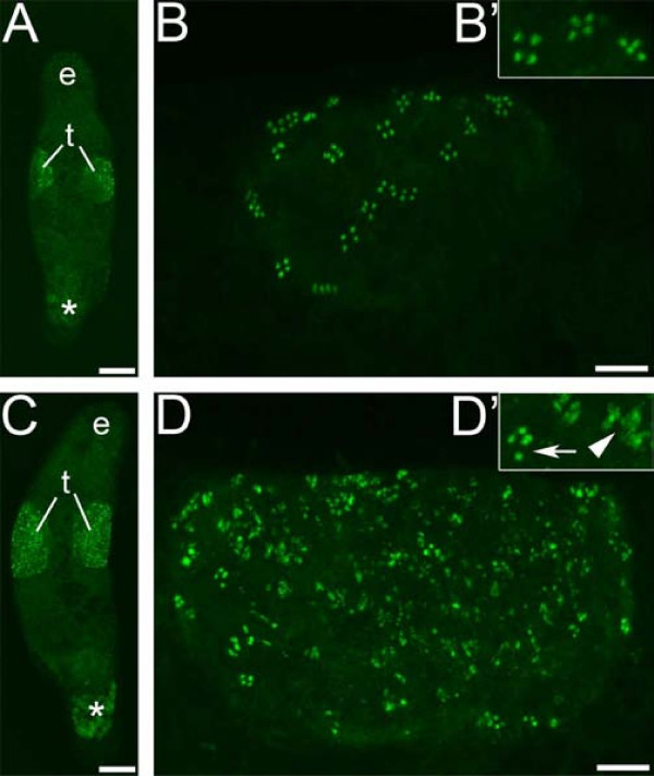
Figure 8.
MSp-1 antibody staining for early spermatid cells in control and melav2 RNAi treated M. lignano. The testis of control animals (A overview, B detail) had normal MSp-1 signal present in clusters of four cells (B, B'), while the testis of melav2 RNAi treated animals (C overview, D detail) had a considerably larger number of MSp-1 positive cells (D, D'). Note that the melav2 RNAi treated animals had some normal MSp-1 signal (D' arrow), but also a lot of disorganized signal (D' arrowhead). e, eyes; t, testes. Asterisk indicates non-specific signal. Scale bars: A, C, 100 μm; B, D, 20 μm.

