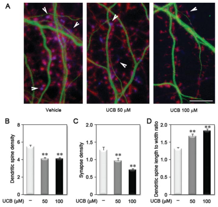
Figure 8.
Treatment of differentiating hippocampal neurons with unconjugated bilirubin (UCB) reduces spinogenesis. A: Representative images of dendrites from neurons treated with vehicle, UCB 50 μM or UCB 100 μM for 24 h at 1 DIV and fixed and labeled at 21 DIV with anti-MAP2 to visualize dendritic shafts (green), phalloidin to visualize actin at the dendritic spines (red), and anti-SV2 to visualize the pre-synaptic terminal (blue). Arrowheads identify a synaptic site by localization of SV2 adjacent to a dendritic spine. Graphics show the effect of UCB on dendritic spine (B) and synapse (C) density, as well as on dendritic spine morphology (D). **p < 0.01 vs. vehicle. Scale bar equals 10 μm. [Color figure can be viewed in the online issue, which is available at www.interscience.wiley.com.]

