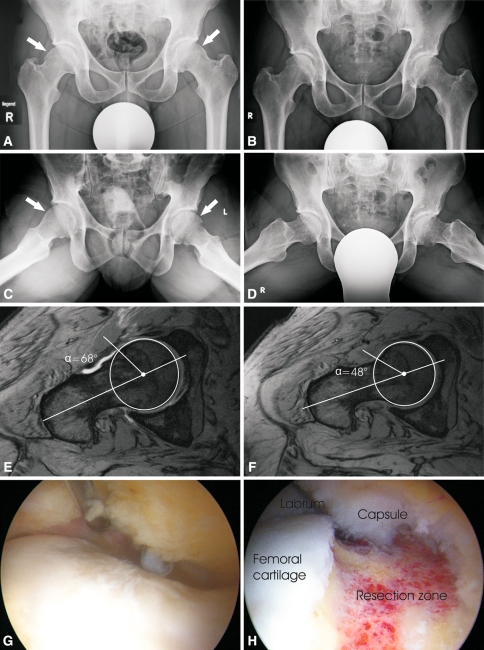
Fig. 3A–H.
The images illustrate the case of a 45-year-old man with symptomatic bilateral femoroacetabular cam impingement. (A) A preoperative AP radiograph of the pelvis and (C) modified Dunn view in 45° flexion show the pathologic bumps at the femoral head/neck junctions (arrows). (B) A postoperative AP radiograph of pelvis and (D) modified Dunn view in 45° flexion show absence of pathologic bumps. (E) A preoperative arthro-MRI shows an alpha angle of 68° (F) whereas a postoperative MRI shows an alpha angle of 48°. Intraoperative photographs show (G) the pathologic bump at the femoral head/neck junction and (H) the femoral head/neck junction after correction of offset.

