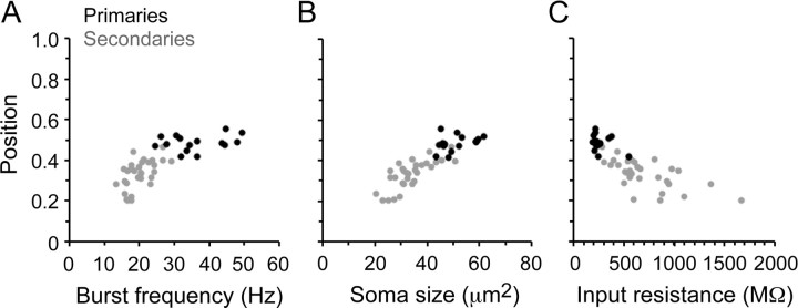
Figure 4.
Motoneuron recruitment, size, and excitability in embryos. A, Plot of spinal cord position versus swimming frequency for 15 primary motoneuron somata (black circles) and 30 secondary motoneurons (gray circles). Correlation values: r = 0.28, p = 0.30, n = 15 (primaries), r = 0.56, p < 0.01, n = 30 (secondaries), r = 0.77, p < 0.0001, n = 45 (combined primaries and secondaries). B, Plot of soma position versus soma size for the same motoneurons in A. Correlation values: r = 0.36, p = 0.19, n = 15 (primaries), r = 0.76, p < 0.0001, n = 30 (secondaries), r = 0.87, p < 0.0001, n = 45 (combined). C, Plot of soma position versus input resistance for the same motoneurons in A and B. Correlation values: r = −0.32, p = 0.25, n = 15 (primaries), r = −0.64, p < 0.001, n = 30 (secondaries), r = −0.79, p < 0.0001, n = 45 (combined). Each data point in A–C represents the mean value for each cell.

