
Fig. 5.
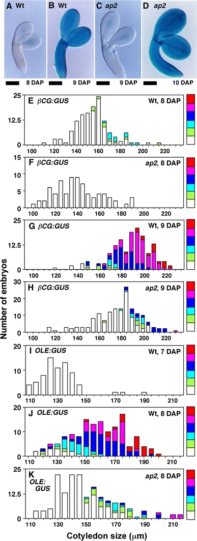
Activation of maturation phase-specific promoters is delayed in ap2 mutants. a–d Wild-type (a, b) and ap2 (c, d) embryos with the β-CG:GUS transgene at the indicated DAP stained for GUS activity. e–h Embryos from crosses between wild-type (e, g, i, j) or ap2 mutant (f, h, k) females and β-CG:GUS (e–h) or OLE:GUS (i–k) males were stained for GUS activity. Histograms show the number of embryos of a given size (indicated by cotyledon width) at a specific DAP. For each size class, the proportion of embryos exhibiting a given intensity of GUS staining is represented, with red indicating the strongest staining and white representing unstained embryos. Unlike wild type, most ap2 embryos containing the βCG:GUS transgene were not strongly stained at 9 DAP, but stained intensely at 10 DAP. Similarly, ap2 embryos containing the OLE:GUS transgene were not strongly stained at 8 DAP, but stained intensely at 9 DAP. Bar = 100 μm