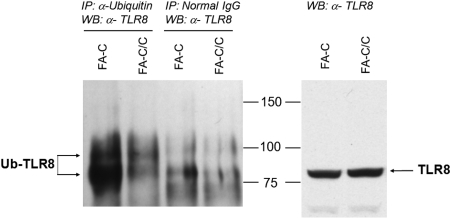
Figure 1.
TLR8-ubiquitin coimmunoprecipitation. Immunoprecipitations were performed by the use of antiubiquitin antibodies (lanes 1-2) or nonspecific control immunoglobulin (lanes 3-4), and immunoblots of the precipitated material were performed by the use of anti-TLR8 antibodies (left). A greater amount of ubiquitinylated TLR8 was detected in immunoprecipitates of mutant cells, confirming the proteomics result. The distinct upper band (lane 1 vs lanes 3-4) is consistent with monoubiquitinylation (8.5 kDa). Total TLR8 protein levels in whole cell lysates were identical in mutant and complemented cells (right). The data shown are representative of 3 identical experiments. A second experiment with loading controls and nonspecific immunoglobulin controls is shown in supplemental Figure 1.

