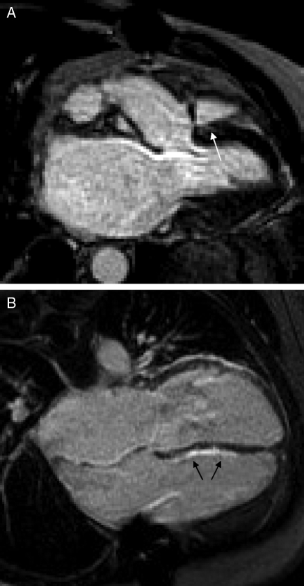
Figure 3.
Late gadolinium-DTPA enhancement images in cardiac transplantation. There is a small area of late gadolinium enhancement in the region of the right and left-ventricular junction of a control patient (A, white arrow). A long-term post-transplant patient with a non-diagnostic endomyocardial biopsy but presumed rejection has a large amount of late gadolinium enhancement in the right-ventricular aspect of the interventricular septum (B, black arrows). The area of bright signal intensity adjacent to the left-ventricular wall clearly extends across the atrioventicular groove, and is likely to represent epicardial fat.

