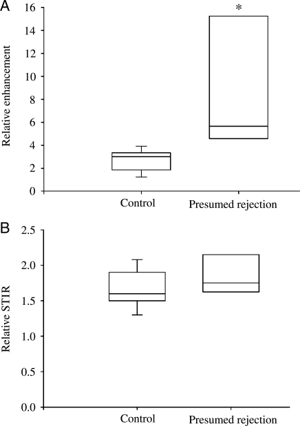
Figure 4.
Cardiac magnetic resonance in presumed rejection. Box and whisker plots of early relative myocardial contrast enhancement (Relative enhancement, A) and myocardial oedema (Relative short inversion time inversion recovery, B) in patients with presumed rejection compared with controls (*P < 0.05).

