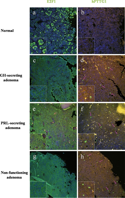
Figure 2.
E2F1 and hPTTG1 immunoreactivity in human pituitary tumor specimens. A and B, Low E2F staining and negative hPTTG1 reactivity in normal pituitary tissue; C–H, high E2F1 and hPTTG1 expression in a nonfunctioning, prolactin (PRL)-secreting and GH-secreting pituitary tumor, respectively. Pink arrows indicate positive hPTTG1 staining cells. Image size, 750 × 750 μm. Green signal, E2F1 or hPTTG1 staining; blue signal, DAPI nuclear staining; yellow signal, autofluorescence background.

