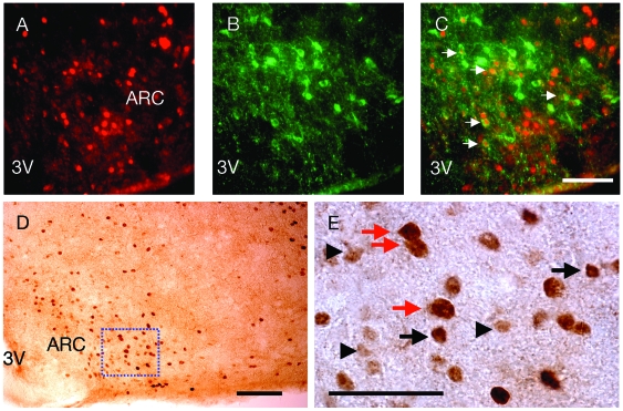
Figure 3. Pancreatic polypeptide (PP) injection induces c-Fos immunoreactivity in neurons positive for alpha melanocyte stimulating hormone (α-MSH) and glutamic acid decarboxylase 65 (GAD65) in the arcuate nucleus of the hypothalamus (ARC).
(A) c-Fos immunoreactivity, (B) α-MSH immunoreactivity, and (C) overlay of c-Fos and α-MSH immunoreactivity in neurons as indicated by white arrows at 30 minutes after i.p. injection of PP. Sale bar for A–C = 25 µm. (D) Brightfield micrograph showing c-Fos and GAD65 immunoreactivity at 30 minutes after i.p. injection of PP. Scale bar = 200 µm. (E) Higher magnification of the boxed area from D. Black arrows indicate neurons positive for c-Fos immunoreactivity only. These neurons are darkly stained. Black arrowheads indicate neurons positive for GAD65 immunoreactivity only. These neurons are lightly stained. Red arrows indicate neurons double-labeled for c-Fos and GAD65. The double staining on these neurons makes these neurons appear larger than the neurons positive only for c-Fos or CAG65 immunoreactvity. Scale bar = 5 µm. 3V, third cerebral ventricle.

