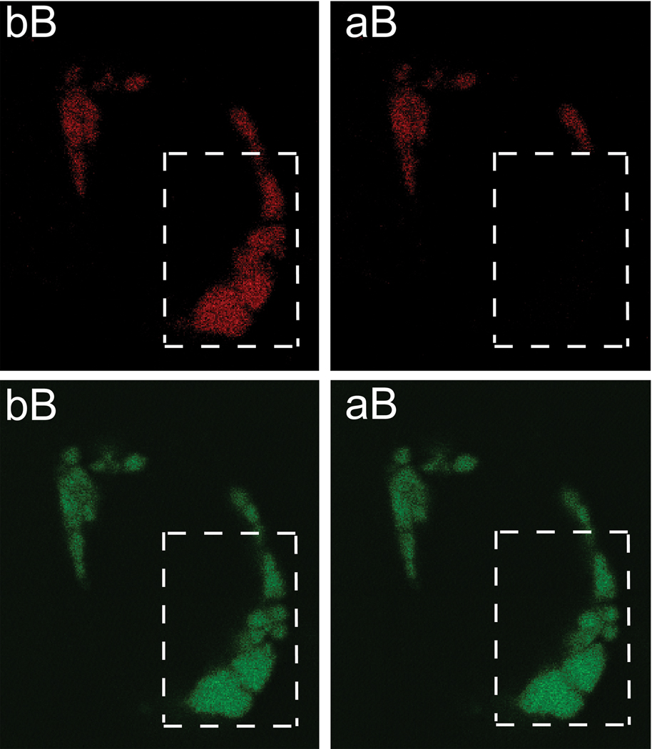
Figure 4.
Representative images of a cell expressing GFP-tagged and RFP-tagged heterogeneous mProVII. The upper panels show images of a cell observed in the red channel while the bottom panels depict the same cell seen in the green channel. Dotted-line boxes indicate an area of a cell subjected to bleaching in the red channel. The relative increase of the green signal after bleaching (aB) in comparison to that before bleaching (bB) indicates occurrence of FRET.

