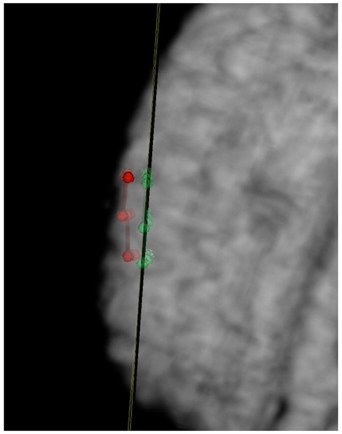
Figure 5. Applications of VVLink to Measure Brain Shift.

Points localized on the surface of the brain using the VVCranial navigational system, whose locations are transferred out via the VVLink interface to the research system for quantification are shown immediately after the craniotomy (in red) and one hour after the craniotomy (in green). An oblique plane is placed showing the average position of these points (in this case one hour post craniotomy) In this case, the brain shift measured was about 5-6 mm.
