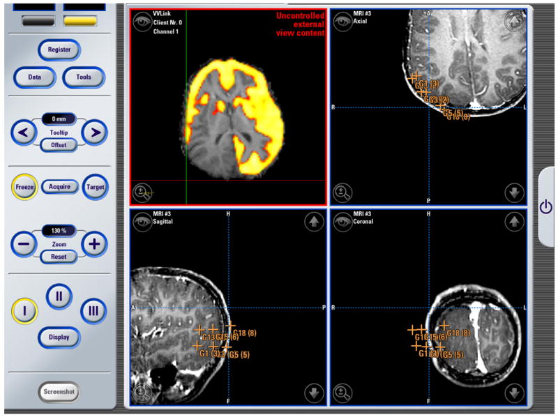
Figure 8. Neurosurgery Example #3.

The image above is a screenshot obtained using the BrainLAB VectorVision Cranial Software during an operation. The top right, and the two bottom views show standard VVCranial visualizations and tool tracking. The top left visualization, which originates from the research system, shows an overlay of a PET image on an anatomical MRI performed using the research workstation and placed into the VVCranial display.
