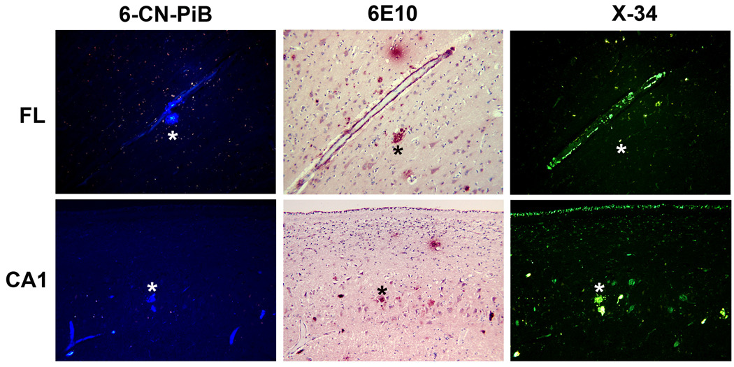
Figure 3.
Fluorescent β-pleated sheet stains label a spectrum of Aβ structures in the frontal lobe (upper panels) and CA1 subfield of hippocampus (lower panels) of postmortem brain of PiBPET-amyloid-negative participant. Amyloid is visible using 6-CN-PiB and X-34, highly fluorescent derivatives of PiB17 and Congo red18, respectively; the monoclonal antibody 6E10, targeting amino acids 1-16 (N-terminus) of Aβ identifies similar structures as denoted by the asterixes.

