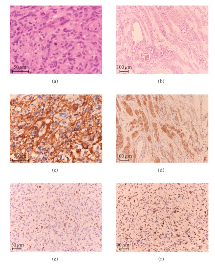
Figure 2.
Histopathological and immunohistochemical findings. Histology of the first lesion showed a cellular astrocytic neoplasm (H&E) (a). The second lesion impressed as a neurofibroma (H&E) (b). The first lesion was GFAP positive (c), whereas the second was not (data not shown). This lesion was however positive for S100 (d). ~10%–15% of the nuclei of the first lesion were immunohistochemically stained for Ki-67 (MiB1) (e). In this lesion, approximately 30% of the cells showed immonupositivity for p53 (f).

