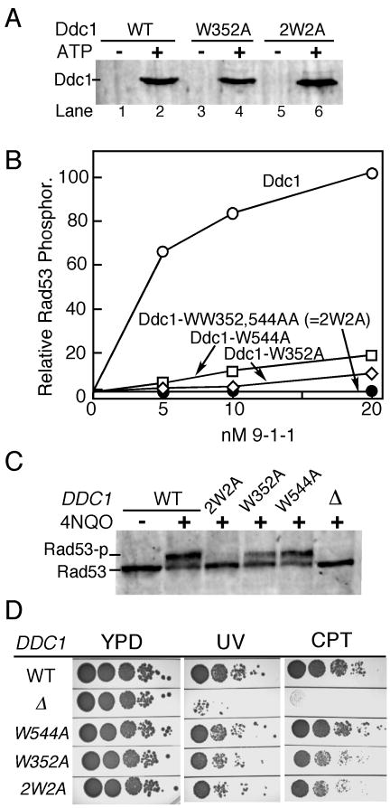
Figure 3. Mapping of bi-partite Mec1 activation motifs in Ddc1.
(A) Ddc1-W352 and Ddc1-2W2A (WW352,544AA) form functional 9-1-1 clamps that can be loaded onto DNA by Rad24-RFC in an ATP dependent manner. See legend to Fig. 1C and Methods for details.
(B) The complete in vitro Mec1 phosphorylation assay was carried out at 125 mM NaCl with indicated levels of (mutant) 9-1-1 clamps (see Fig 1D and Methods for details). Phosphorylation of Rad53-kd is quantified. Background phosphorylation of Rad53-kd by Mec1, obtained in the absence of Ddc1 was substracted.
(C) Western analysis of G1-arrested cells exposed to 4NQO for 30 min.
(D) Ten-fold serial dilutions of wild type cells and the indicated DDC1 mutants were tested for sensitivity to UV (60 J/m2) or camptothecin (10 μg/ml). Plates were incubated at 30 °C for two days and photographed.

