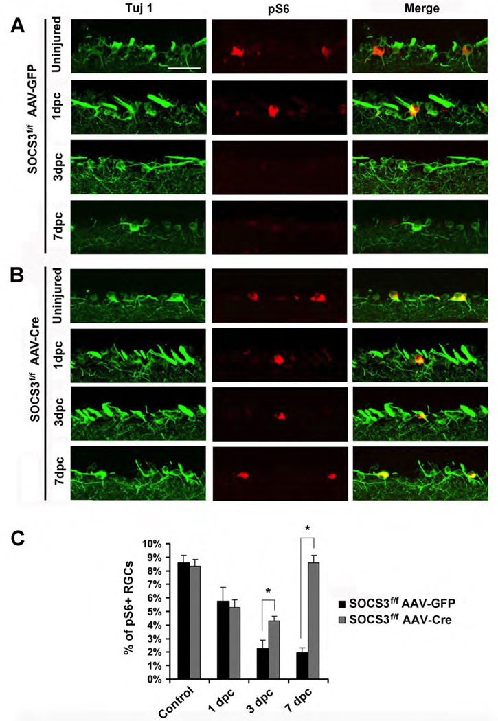
Figure 2. Phospho-S6 levels in RGCs of SOCS3f/f mice with AAV-GFP or AAV-Cre after optic nerve injury.
(A, B) Immunofluorescence analysis with anti-p-S6 or TUJ-1 antibodies of the retinal sections from SOCS3f/f mice injected with AAV-GFP (A) or AAV-Cre (B) at different time points post-crush. Scale bar, 50 µm.
(C) Quantification of p-S6+ RGCs. Data is presented as mean percentages of p-S6+and TUJ1+ cells among total TUJ1+ cells in the ganglion cell layer of each retina. Cell counts were performed on at least 4 non-consecutive sections for each animal, from four or five mice per group. *: p<0.01 by Dunnett’s test.

