
Figure 3.
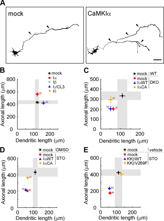
A specific role for a CaMKK–CaMKIα cascade in promoting axonal growth in cortical neurons. A, Representative images of rat cortical neurons transfected with GFP-CaMKIα. Scale bar, 50 μm. B, Overexpression of CaMKIα and CaMKIγ facilitated axonal and dendritic growth, respectively. n = 15 for all groups. aAxon, p < 0.001; bdendrite, p < 0.05 (one-way ANOVA with Tukey's test comparison with mock). C, The axonal growth defect in DKO mice was selectively rescued by coexpression of a constitutively active CaMKIα (CaMKIαCA), but not by a wild-type CaMKIα (CaMKIαWT); the dendritic growth defect was left unaltered; n = 15 for all groups. aAxon, p < 0.01; dendrite, p < 0.05; baxon, p < 0.01; dendrite, p < 0.001; cdendrite, p < 0.01 (one-way ANOVA with Tukey's test comparison with WT plus mock). D, Only the axonal growth defects caused by STO-609 treatment were rescued by expression of a constitutively active CaMKIα (CaMKIαCA), but not of a wild-type CaMKIα (CaMKIαWT). Dendritic growth defects remained unchanged; n = 15 for all groups. a,bAxon, p < 0.001; dendrite, p < 0.001; cDendrite, p < 0.001 (one-way ANOVA with Tukey's test comparison with DMSO plus mock). E, Both axonal and dendritic growth defects caused by STO-609 treatment were rescued by introducing an STO-609-resistant CaMKKβ mutant (V269F), but not a CaMKKβ-WT. n = 15 for all groups. a,bAxon, p < 0.001; dendrite, p < 0.01 (one-way ANOVA with Tukey's test comparison with vehicle plus mock).