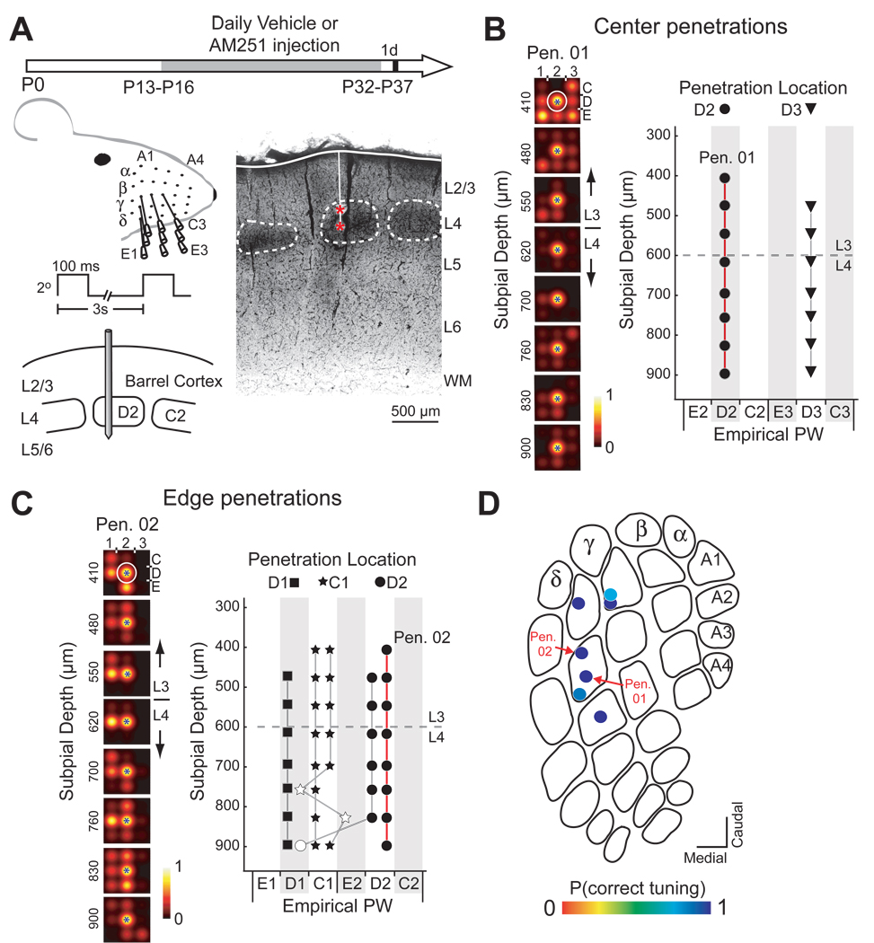
Figure 1. Whisker map precision in a vehicle-treated rat.
(A) Left: Experimental design and electrophysiological recording. Right: Coronal section showing a penetration with two marking lesions (red *). Scale bar: 500 µm. (B) Multiunit receptive fields (RFs) for recording sites along a penetration through the D2 column center (PN01) in rat H46. Circle, anatomically corresponding whisker for the penetration. Blue star, measured empirical PW for each recording site. (C) Empirical PW at all recording sites in 2 center penetrations in H46. Symbol shape denotes recording site location and anatomically appropriate whisker. Filled symbols, correctly tuned sites. Dashed line, L2/3-L4 border. (D) and (E) Tuning curves and PW measured for all 5 edge penetrations in H46. Open symbols denote mistuned sites. (F) P(correct tuning) for all penetrations in H46, plotted on an exemplar barrel map.

