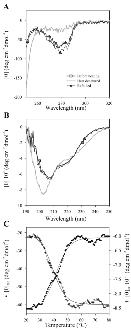
Fig 2.
Reversible heat denaturation of PVA VPg monitored by CD spectroscopy. Spectra were measured at +20 °C, at +80 °C, and after heat denaturation followed by renaturation at +20 °C. The sample concentration was 0.2 mg/ml. 5 scans were added and averaged. Temperature dependence of PVA VPg CD spectra was followed (A) in near-UV region (250–320 nm), and (B) in far-UV region (190–260 nm). (C) Thermal unfolding curves were measured at 203 nm and at 280 nm with a constant heating rate of 1°C/min. The data are given as mean molar ellipticity per residue at 280 nm (●; scale on the left) and at 203 nm (+; scale on the right) plotted against increasing temperature. The melting temperature of PVA VPg structure is around 42°C. Nearly complete reversibility after heat treatment was observed.

