Figure 2.
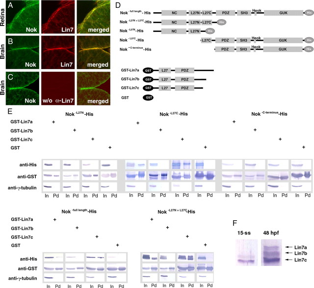
Lin7c is expressed during neurulation and physically interacts with the L27C domain of Nok. A–C, Sequential labeling of Lin7 (red) and Nok (green) revealed their colocalization to the apical surface of the retina (A) and the brain epithelia (B) at 24 hpf. No red fluorescence was detectable when pan-Lin7 primary antibodies were not used (C), demonstrating the specificity of the sequential labeling. D, The diagram illustrates the constructs of the Nok-His and GST-Lin7 fusion proteins that were used in the pulldown analysis (E). E, A GST pulldown analysis demonstrated that Nok interacts with Lin7c and Lin7a via its L27C domain. Anti-γ-tubulin blots confirmed that the His signals in the pulldown fractions were not due to nonspecific precipitation. In, Input fractions; Pd, pulldown fractions. F, A Western blot analysis revealed that Lin7a is not detectable at the 15-ss and that Lin7c is expressed at a much higher level than Lin7b. In contrast, all three Lin7 genes are expressed at 48 hpf.

