Figure 5.
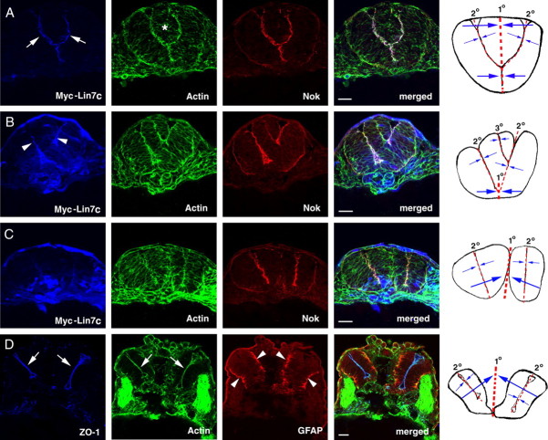
Overexpression of Lin7c induces either multiaxial mirror symmetry in the CNS or multiple neural tubes. The embryos injected with Myc-Lin7c mRNA were examined at the 20-ss or 30 hpf for neural tube development and for the localization of basal maker GFAP and apical markers ZO-1, Nok, and actin bundles. The column at right is the schematic drawings that illustrate the multiaxial mirror symmetry of the CNS. Red-dashed lines mark the axes of mirror symmetry. 1° indicates primary axes; 2° indicates secondary axes; and 3° indicates ternary axes. Opposing blue arrows illustrate the mirror arrangements of cellular and subcellular structures. A–C, When moderately expressed, Myc-Lin7c (blue) localized apically at the bifurcated apical surface (A) at the 20-ss. The asterisk indicates the accumulation of cells between the branches of the bifurcated apical surface. When expressed at higher levels (B, C), Myc-Lin7c localized both apically (arrowheads) and diffusely at the 20-ss. In some embryos, bifurcated multiaxial mirror symmetry was observed (B); in other embryos, two or more separate neural tubes were present (C). D, At 30 hpf, basal marker GFAP (red, arrowheads), apical marker ZO-1 (blue, arrows), and actin bundles (green, arrows) localized to the proper regions in each separated neural tube. Scale bars, 30 μm.

