
Figure 7.
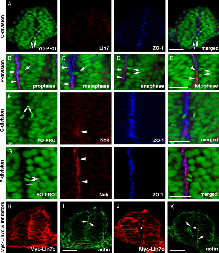
Temporal and spatial regulation of the expression of Lin7c complex during C- and P-division is critical for proper neural tube development. M-phase nuclei were identified by the condensed chromatin morphology (A–G, YO-PRO staining in green). A, Lin7c (red) is undetectable in the neural rod regions that exhibit C-division at the 15-ss. Arrows indicate the sister-daughter nuclei at the anaphase/telophase, one on each side of the midline region marked by the ZO-1 staining (blue). B–E, During P-division, Lin7c (red, arrowheads) is strongly present at the apical surface of the neuroepithelium, where ZO-1 (blue) localizes. Arrows indicate P-division nuclei at prophase (B), metaphase (C), anaphase (D), and telophase (E). No Lin7c is observed at the cleavage furrow during anaphase and telophase (D, E). F, Nok expression is also absent in regions undergoing C-division (arrows) at the 15-ss. The ZO-1 staining (blue) marks the midline region. However, ventral to the C-division nuclei, Nok signal was apparent at the ventral-most midline region in the neural rod (arrowhead). G, At the 20-ss, strong Nok staining (red, arrowheads) was present at the apical surface of the neuroepithelium where a P-division (arrows) was occurring. H–K, In the presence of cell division inhibitors, overexpression of Myc-Lin7c (red, stained with anti-Myc antibody) did not induce multiaxial mirror symmetry at either the neural rod stage (H, I) or the neural tube stage (J, K). Actin (green) accumulation at the apical surface of the neuroepithelium (arrows) confirms the presence of single-axial mirror symmetry. Asterisks indicate the lumen of the neural tube (J, K). Scale bars, 30 μm.