
Figure 8.
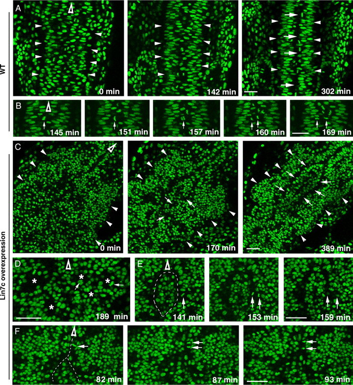
Overexpression of Lin7c causes abnormal cell movements and cell–cell adhesion, which lead to the formation of multiple neural tubes. The movements of nuclei in H2A-GFP transgenic embryos (Pauls et al., 2001) were examined with live confocal imaging in the coronal plane, starting at the 12-ss. Hollow arrowheads indicate the anterior direction. A, Time-lapse images of the neural keel-to-rod transition (started at the 10-ss) reveal the narrowing of the tissue and the gradual formation of the midline region marked by the interphase-nucleus-free zone (arrows) in wild type. Arrowheads indicate the lateral boundaries of the CNS. B, Time-lapse imaging reveals the midline crossing of one of the daughter cells (arrows) during a C-division in wild type. C, Time-lapse images revealed the irregular CNS development in an embryo that overexpressed Myc-Lin7c. Arrows indicate that the interior regions of the cellular rosettes gradually fused to realign in an anterior–posterior direction. At 389 min, two anterior–posterior-aligned interphase-nucleus-free zones (arrows) are evident. Arrowheads indicate the lateral boundaries of the CNS. D, M-phase nuclei (arrows) were positioned in the interior (asterisks) of cellular rosettes, which were randomly packed in the CNS. E, Time-lapse imaging revealed a cell division that resembles the C-division of wild-type embryos. However, the crossing daughter cell got stuck with a group of cells in the middle of the CNS. This group of cells was isolated by two interphase-nucleus-free zones (dashed line). F, At the regional anterior–posterior-aligned apical surfaces, a significant number of cell divisions resembled P-divisions (arrows indicate one example). The dashed line indicates the interphase-nucleus-free zone. Scale bars, 30 μm.