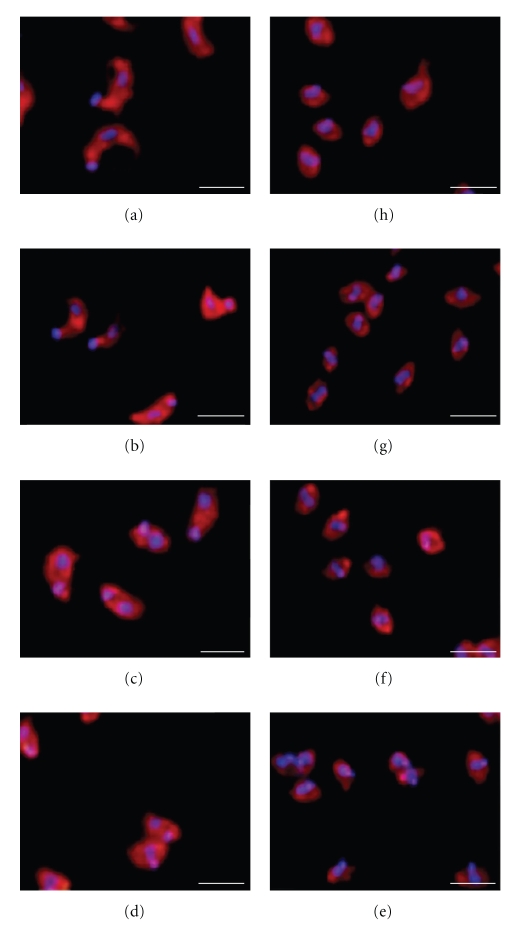
Figure 3.
The shape and position of the nucleus and the kinetoplast identified the different IFs. The morphology and position of the nucleus and the kinetoplast were determined by indirect immunofluorescence and DAPI staining of trypomastigotes (a), IFs at 1 hour (b), 2 hours (c), 3 hours (d), 4 hours (e), 5 hours (f) and 6 hours (g) of transformation and amastigotes (h). Bar = 25 μm.

