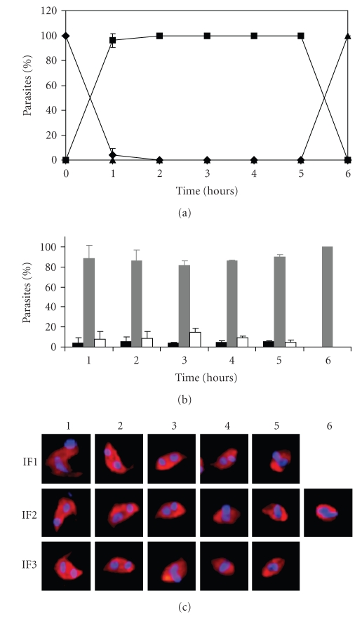
Figure 4.
Highly synchronous morphologic changes are obtained during the in vitro transformation kinetics. Quantitative analysis of 2 independent experiments of extracellular differentiation from tissue-derived trypomastigotes into amastigotes stained by indirect immunofluorescence and DAPI after 1, 2, 3, 4, 5 and 6 hours of transformation. (a) Relative percentage of trypomastigotes (-♦-), IFs (-■-) and amastigotes (-▲-). (b) Three different IFs arbitrarily designated as IF1 (black), IF2 (grey) and IF3 (white) were observed from 1 to 5 hours of transformation. (c) Representative parasite of the corresponding IFs of panel b.

