
Figure 2. Digital images from the electron microscopy of TG and peritubular capillary laminations.
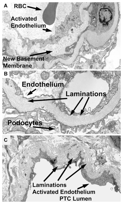
(A) Glomerular loop with new basement membrane (arrow), that is duplication, activated endothelium (arrow) and lumen with a red blood cell (RBC, arrow). Original magnification 7100×. (B) Glomerular loop with thickened basement membrane laminations (arrow), and activated endothelium (arrow). Podocytes for reference (arrow). Original magnification, 15 000×. (C) Peritubular capillary with laminations (arrow). and activated endothelium (arrow). Peritubular capillary lumen for reference (PTC lumen). Original magnification, 9100×.