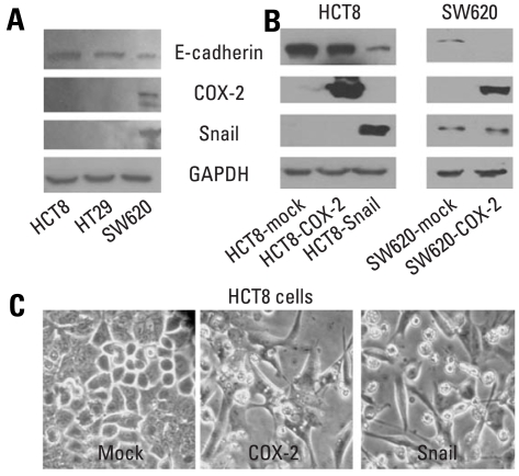
Fig. 1.
Western blot analyses of E-cadherin, COX-2 and Snail in colon cancer cells, and morphology of HCT8 transfected with cDNA for COX-2 or Snail. (A and B) Endogenous expressions of E-cadherin, COX-2, and Snail in colon cancer cells, and ectopic expressions of COX-2 and Snail in HCT8 and SW620. Forty µg of protein was separated by 10% SDS-polyacrylamide gel electrophoresis and transferred to a nitrocellulose membrane. The bottom represents GAPDH, which was used as a loading control. (C) Ectopic expression of COX-2 or Snail induced a scattered, flattened phenotype with few intercellular contacts in HCT8. COX-2, cyclooxygenase-2; SDS, sodium dodecyl sulfate; cDNA, complementary DNA; GAPDH, glyceraldehydes 3-phosphate dehydrogenase.

