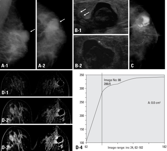
Fig. 3.
A 39-year-old woman presented with bloody nipple discharge on her left breast. (A) Left mammography, MLO (1) and CC (2) view showed heterogeneous dense parenchyma pattern and well-circumscribed high density mass lesion at left upper outer portion of breast (white arrow). No evidence of microcalcifications. (B) Breast ultrasonography (a) showed 2.0 × 1.7 cm sized mass lesion at left 2 : 00 portion. The mass was complex cystic mass with internal lobulated hypoechoic solid lesion. Focal ductal extenstion toward the nipple was noted (white arrows). On Doppler image, peripheral increased blood flow was seen. There was no blood flow within the mass (b). (C) On galactography, dilated nipple duct was noted and contrast dye which filled the cystic portion of the mass lesion and solid lesion demonstrated to be lobulated filling defect. (D) Dynamic contrast breast MRI subtraction 1 minute (1), 3 minutes (2), and 5 minutes (3) images. The mass lesion at left 2 : 00 showed peripheral thin rim enhancement. The solid portion of the mass did not reveal enhancement in dynamic images. The enhancement kinetics demonstrated persistent pattern (4), therefore, the BI-RADS category was 3. The pathologic result after excision was intraductal papilloma.

