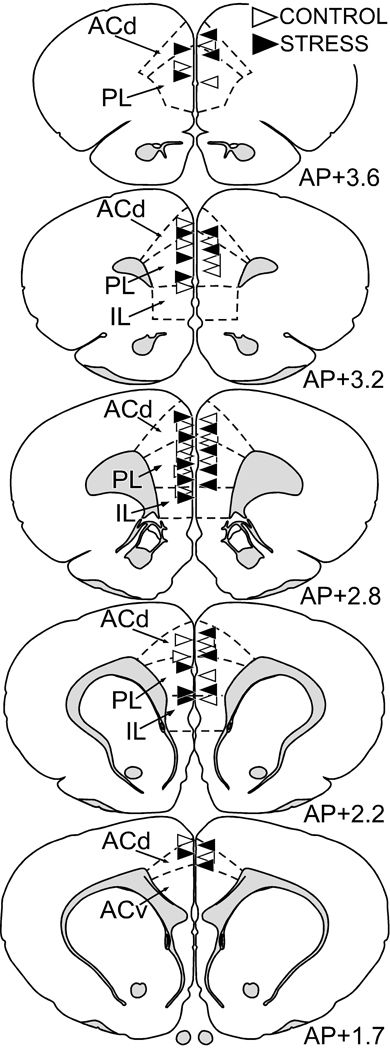
Figure 3. Anatomical localization of Lucifer Yellow-filled layers II/III pyramidal neurons in mPFC for subregional analysis.
Atlas plates (modified from Swanson, 1992) of coronal sections through a similar level of the mPFC from animals that received intracellular Lucifer Yellow injections. Neurons from layers II/III in the dorsal mPFC were identified using a fluorescent nucleic acid stain, followed by iontophoretic injections with Lucifer Yellow. The location for each filled neuron is designated for each treatment with a triangle (control, blue; stress, red). ACd, anterior cingulate cortex, dorsal subdivision; ACv anterior cingulate cortex, ventral subdivision; PL, prelimbic area; IL, infralimbic area.

