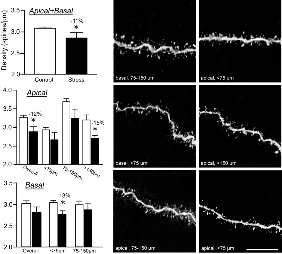
Figure 4. Repeated restraint stress reduces dendritic spine density in layer II/III pyramidal neurons.
Repeated restraint stress (21 days, 6 hours/day) induces decreases in overall (upper left) and apical dendritic spine density (middle left). This effect is most prominent in more distal portions (>150 µm) of apical dendrites. While there was no overall effect of stress on basal dendrites, a significant reduction of spine density was present at <75 µm from the soma (lower left). The spine density values for each dendritic segment were quantified using the Rayburst-based automated approach and was similar to previous estimates that involved manually counting spines from optical stacks by controlling the plane of focus for z-step increments and marking spines as they appear on the dendritic segment (Radley et al., 2006b). Examples of dendritic segments are shown in the middle (control) and right (21 days stress) columns. *, differs significantly from unstressed controls; P < 0.05. Scale bar = 10 µm.

