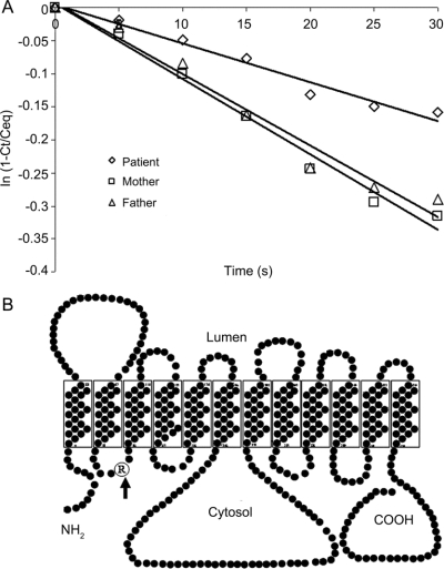
Figure Glut1 protein: Functional and structural analysis
(A) 3-O-methyl-d-glucose uptake into red blood cells, which was performed as described elsewhere.4 Decreased uptake rate in patient is shown compared with parents serving as control. The data are expressed as the natural logarithm of the ratio of intracellular radioactivity at time T/equilibrium vs time in seconds (2 determinations/data point). (B) The proposed Glut1 protein configuration with the R93W mutation (arrow) situated in the first cytosolic loop.

