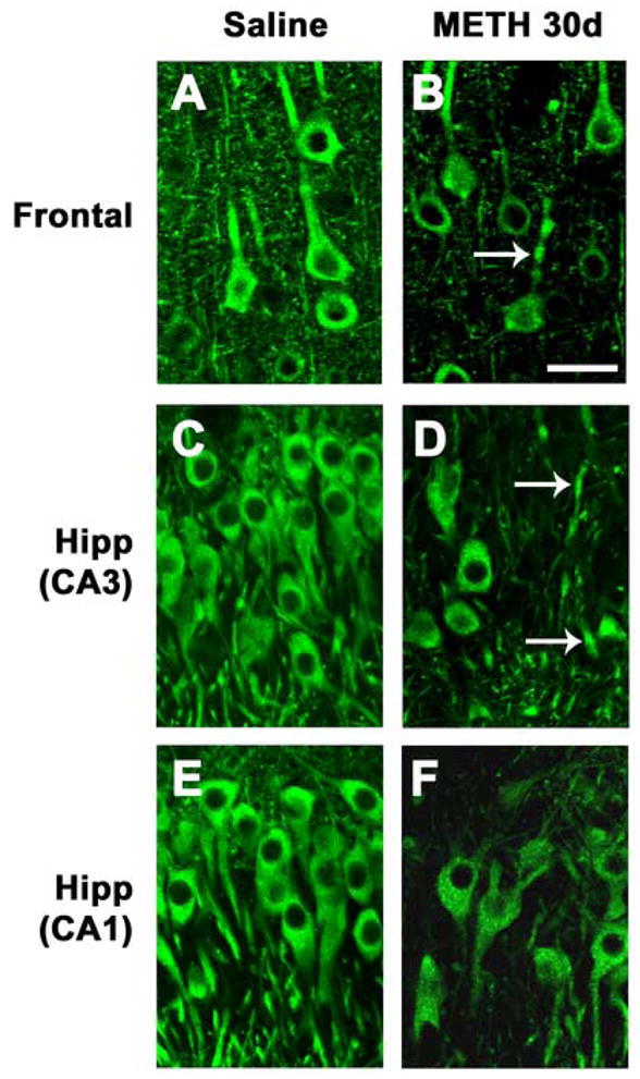
Fig. 4.

Dendritic pathology in the neocortex and hippocampus after METH binge treatment. Sections were immunolabeled with an antibody against MAP2 and imaged with the laser scanning confocal microscope. (A) Preserved dendritic architecture of frontal cortex neurons in a saline-treated rat. (B) 30d post METH binge, neurons in the frontal cortex are shrunken and some have dystrophic neurites (arrow). (C) Preserved dendritic architecture of neurons in the CA3 region of the hippocampus in a saline-treated rat. (D) 30d post METH binge, pyramidal CA3 neurons are shrunken with tortuous processes (arrows). (E) Preserved dendritic architecture of neurons in the CA1 region of the hippocampus in a saline-treated rat. (F) 30d post METH binge, pyramidal CA1 neurons are shrunken with tortuous processes. Scale bar, 20μM. *p<0.05 compared to saline-treated controls by one-way ANOVA with post hoc Dunnett’s.
