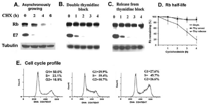
Figure 1.
Rb proteolysis is blocked in the S-phase of Caski cells. Caski cells were arrested in S-phase by double thymidine block as described in the materials and methods. Asynchronously growing cells (panel A), thymidine arrested cells (panel B), and thymidine-released cells (panel C) were treated with cycloheximide (25ug/ml) for the indicated time. Cell lysates were analyzed for Rb, E7, and α-tubulin (loading control) using the Western blot assay. (D). A quantification of the Rb band intensity was plotted against the time after the cycloheximide addition. The average of two independent experiments is shown. (E) Cell cycle profiles of asynchronous, thymidine arrested and thymidine released Caski cells are shown.

