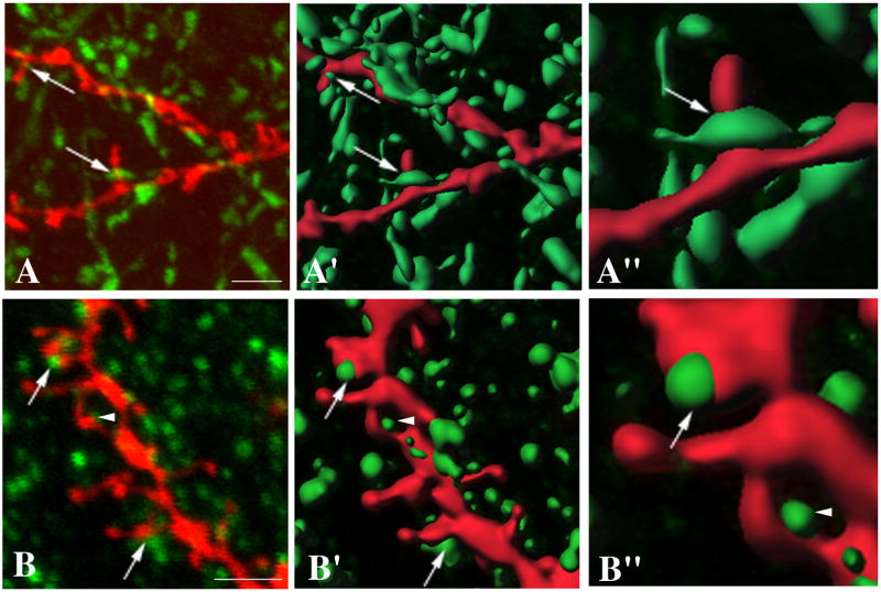
Figure 2. Combination of diolistic labeling with immunofluorescence.
A. MSNs in the striatum of triple organotypic cultures were diOlistically labeled with CM-DiI (red) and the TH-fibers visualized by immunofluorescence (green). Areas of apposition between TH-ir afferents and the necks of MSN spines (arrows) were determined using Bitplane software. B. Organotypic triple cultures were labeled with CM-DiI (red) and immunocytochemically stained using anti-VGluT1 antibodies (green). Areas of apposition between MSNs dendritic spine heads and VGluT1-ir terminals were determined using Bitplane software (arrows). A VGluT1-ir presynaptic terminal that appeared close, but did not colocalize with the CM-DiI labeled dendritic spine is indicated by an arrow head. A′,B′, A″, B″. Three-dimensional reconstructions are rendered using Bitplane software. Scale bar = 2.5 μm in A,A′ and B,B′. A″ and B″ show 2.5-fold magnified examples of TH-ir and VGlut1-ir appositions with CM-diI labeled MSNs spines respectively.

