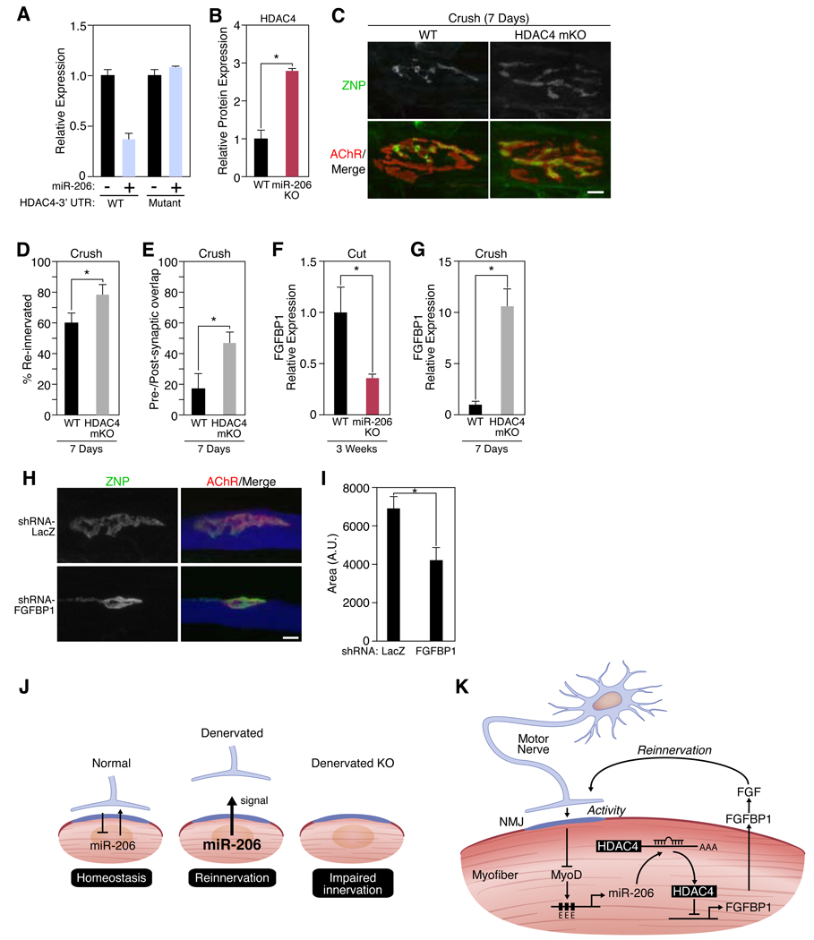
Figure 4. miR-206 targets HDAC4.
(A) Luciferase activity of COS1 cells co-transfected with WT or mutant HDAC4 3’UTR-luciferase constructs with a miR-206 expression plasmid. (B) Quantitation of HDAC4 protein expression in muscle lysates isolated from wild-type (WT) and miR-206−/− (miR-206 KO) mice following 3 weeks of denervation. *p < 0.02 by t test. n=3. (C) Immunohistochemistry shows an increase in reinnervation in HDAC4 mKO mutant mice compared to WT mice 7 days following nerve crush. Scale bar=10 µm. (D) Number of reinnervated NMJs in WT and HDAC4 mKO mice following sciatic nerve crush for 7 days. *p < 0.05 by t test. n=3–8. (E) Postsynaptic area occupied by the reinnervating nerve (in %) 7 days after crushing the sciatic nerve in WT and HDAC4 mKO mice. *p < 0.02. (F) Decrease in expression of Fgfbp1 transcripts in miR-206−/− (KO) muscles 3 weeks after nerve transection. *p < 0.02 by t test. n=3–5. (G) Increase in expression of Fgfbp1 transcripts in HDAC4 mKO muscles 7 days after nerve crush. *p < 0.0005 by t test. n=3–5. (H) Immunohistochemistry shows an inhibition of synaptic-vesicle clustering in neonatal NMJs upon knock-down of FGFBP1. Scale bar= 10 µm. (I) NMJ size in muscle fibers expressing LacZ or FGFBP1 shRNAs. Mice were electroporated at P0 and analyzed at P8. *p < 0.02 by t test. n=4. Values represent means ± SEM (J) Schematic of miR-206 up-regulation and function after denervation. (K) Mechanism of miR-206-dependent reinnervation.

