Figure 15.

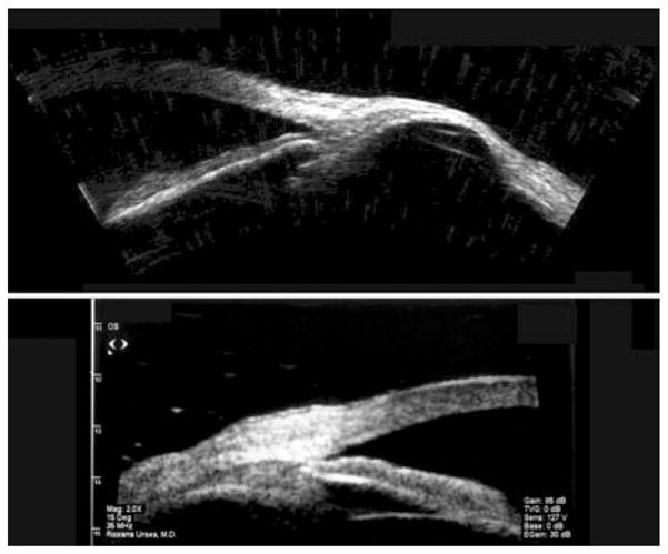
Top: Scleromalacia 6 years following ruptured globe. Image acquired using Cornell prototype scanner. Bottom: Thickened sclera in localized anterior uveitis, demonstrated with Sonomed UBM. (Image courtesy of Roxana Ursea, MD.)

Top: Scleromalacia 6 years following ruptured globe. Image acquired using Cornell prototype scanner. Bottom: Thickened sclera in localized anterior uveitis, demonstrated with Sonomed UBM. (Image courtesy of Roxana Ursea, MD.)