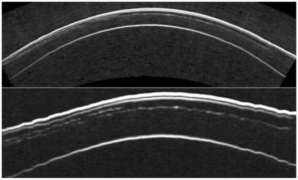
Figure 8.
An incomplete flap occurred during the primary LASIK treatment of this eye because of the patient’s squeezing during the Hansatome microkeratome pass. Two months later, the patient was scanned using the Artemis-1 and the maximum flap thickness was measured to be 176 μm. The patient was then retreated using the VisuMax femtosecond laser (Carl Zeiss Meditec, Jena, Germany) with a programmed flap thickness of 190 μm so that the second flap would be deeper than the first flap. The images, top in geometrically correct format, bottom in stretched rectilinear format, show the cornea in the horizontal meridian after the second procedure. Both the initial, incomplete Hansatome flap and the complete VisuMax flap interfaces can be clearly seen. (Image courtesy of Dan Z. Reinstein, MD MA[Cantab] FRCSC.) LASIK, laser in situ keratomileusis.

