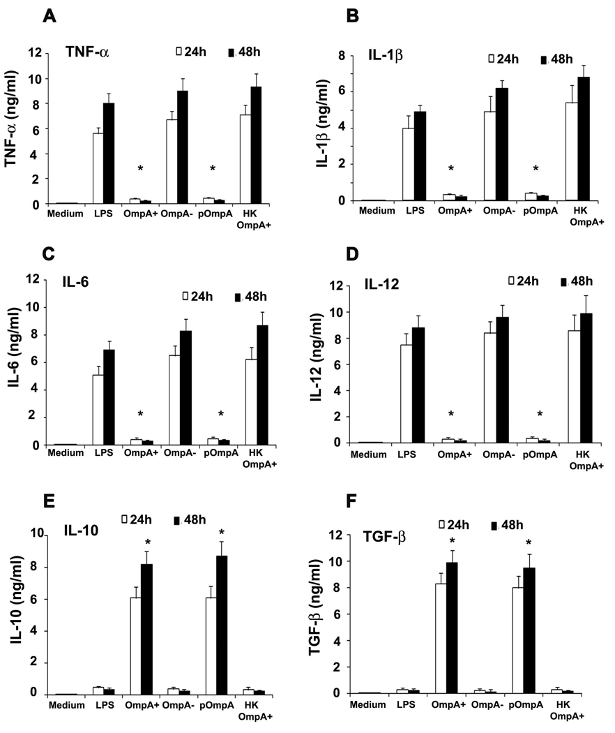
Figure 3. Cytokine production by DCs infected with ES.
DCs were exposed to medium alone, treated with various ES strains, LPS or heat killed (HK) ES for 24 and 48 h. Culture supernatants were collected and the levels of TNF-α (A), IL-1β (B), IL-6 (C), IL-10 (D), IL-12p70 (E), and TGF-β (F) were assessed by ELISA. The error bars represent standard deviations from the means of triplicate samples from four individual experiments. The suppression of cytokine production by OmpA+ or pOmpA+ ES was significant in comparison to OmpA− ES, LPS or HK ES induced levels, *p<0.001 by two tailed t test.

