


Fig 1.
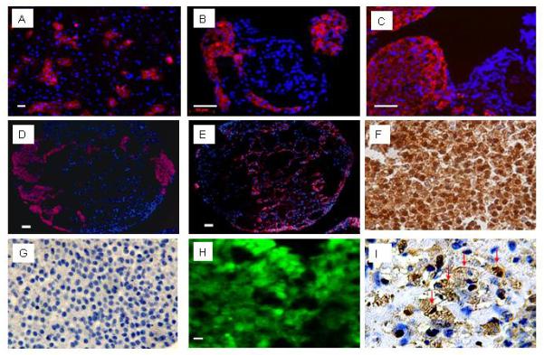
Characterization of hESC-derived cardiomyocytes. A-E: Marker gene expression was demonstrated using rhodamine-conjugated secondary antibodies. Panel A shows anti-alpha-actinin immune reactivity (red) to a monolayer of hESC-derived cells isolated via trypsin-digestions of spontaneously contracting EBs. B-E: Immune staining to EB sections. Panel B shows anti-αMHC immune reactivity, Panel C shows anti-tropomyosin immune reactivity. Serial sections show anti-MLC2a immune reactivity in panel D, and cytoceratin 8 immune reactivity in panel E. Hoechst staining was employed to identify nuclei (blue signal); scale bar, 50 μm. F: Immune staining serial sections from a hESC-derived EB with antibody against GFP (HRP-conjugated secondary antibody, signal developed with diaminobenzidine reaction) (400×). G: negative control for the anti-GFP staining (i.e., without primary antibody against GFP) (400×). H: Epi-fluorescence signal in a hESC-derived EB section (10 μM thick). I: Anti-sarcomeric actin immune reactivity in a hESC-derived EB section (HRP-conjugated secondary antibody, signal developed with diaminobenzidine reaction). Many cells are positive to sarcomeric actin (brown) and display cross striations which appear dark brown (marked by red arrows; 600×).