


Fig 10.
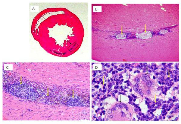
H&E staining of Sprague-Dawley rat heart at 4 weeks after cell injection. A: hematoxylinophilic staining (blue) demarks the area where cells were delivered to the heart. B: higher magnification of the boxed area indicted by black arrow in panel A; overt necrosis in the engrafted area surrounded by lymphocytic infiltrate is apparant (yellow arrows, 200×). C: higher magnification of the boxed area indicted by white arrow in panel A; the lymphocytic infiltrate in the graft area is apparent (yellow arrows, 200×). D: higher magnification of the boxed area in panel C; giant cells (black arrows) are surrounded by the lymphocytic infiltrate (yellow arrows) (400×).