
Fig 3.
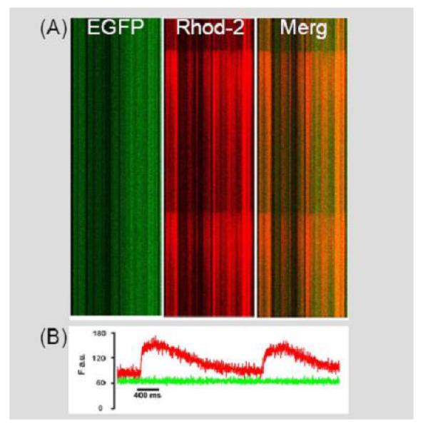
Shipped hESC-derived EBs exhibit electric field-stimulated intracellular calcium transients. A: Stacked line scan images depicting electrically evoked calcium-dependent increases in rhod-2 fluorescence in cultured hESC-derived EBs (y-axis, time; x-axis, position). The left panel shows the EFGP epi-fluorescence signal and the middle panel shows the rhod-2 fluorescence signal. The signals were merged in the right panel. B: Spatially integrated traces of the changes in rhod-2 (red) and GFP (green) fluorescence during field stimulation-induced depolarizations in hESC-derived EBs.