
Fig 4.
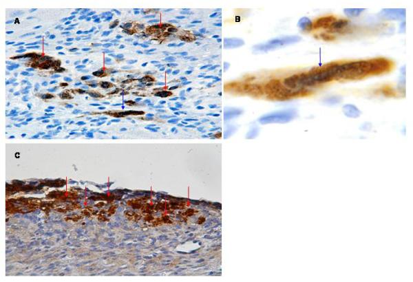
Detection of hESC-derived cardiomyocytes following transplantation into ischemic myocardium. A: Anti-GFP immune reactivity of a heart examined 1 week following cell transplantation (HRP-conjugated secondary antibody, signal developed with diaminobenzidine reaction; see red arrows) (400×). B: Higher magnification of cell indicated by blue arrow in panel A. The cell shows cross striations (blue arrow) and demonstrates a more cylindrical shape (600×). C: Anti-GFP immune reactivity of a heart examined 4 weeks following cell transplantation, processed as described in panel A (400×).