
Fig 5.
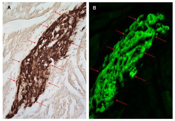
Co-localization of GFP immune reactivity and epi-fluorescence in serial sections from a heart receiving hESC-derived cardiomyocytes (2.5 hours post-transplantation). A: Anti-GFP immune reactivity (HRP-conjugated secondary antibody, signal developed with diaminobenzidine reaction; see red arrows). B: GFP epi-fluorescence in an adjacent section to that depicted in panel A.