
Fig 7.
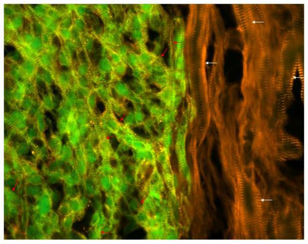
A merged image with GFP epi-fluorescence (green) and alpha-actinin immune reactivity (red, rhodamine-conjugated secondary antibody) to show the juxtaposition of hESC-derived donor cardiomyocytes and the host myocardium at 4 weeks after transplantation in nude rat. Donor cells are green with yellow color cross striations (red arrows). Host cells show red color cross striations (white arrows). Note that the diameter of transplanted cells (green) is smaller than native cells, and there is some myofiber disarray in the transplant (red arrows).